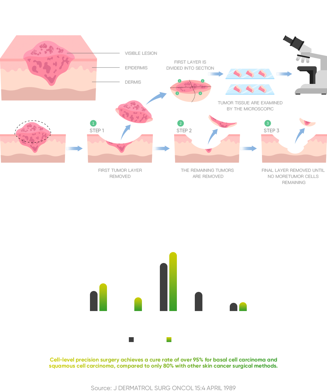


In 1936, Dr. Frederic Mohs discovered that examining tumor margins with a microscope and removing tissue layer by layer during surgery could maximize tumor cell removal while preserving normal tissue. This technique increased the cure rates for basal cell and squamous cell carcinomas from 80% to over 95%. However, the complexity and length of Mohs surgery have limited its use to other cancers for over 80 years.
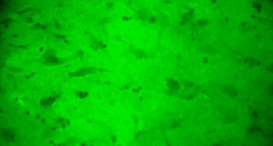
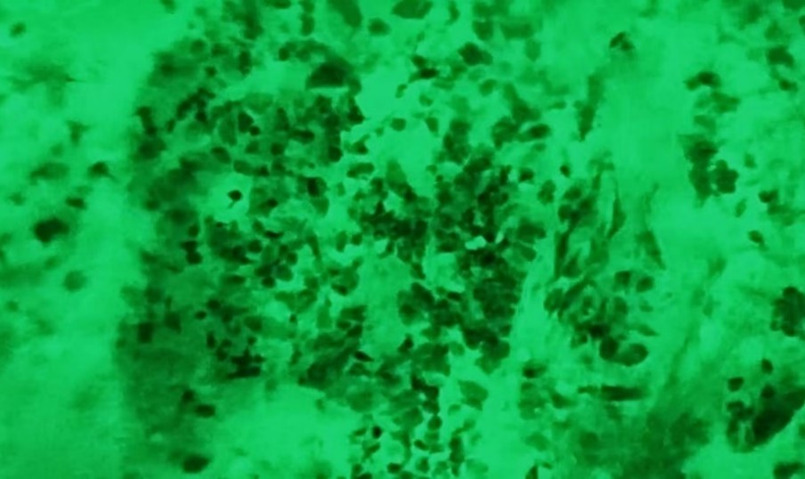
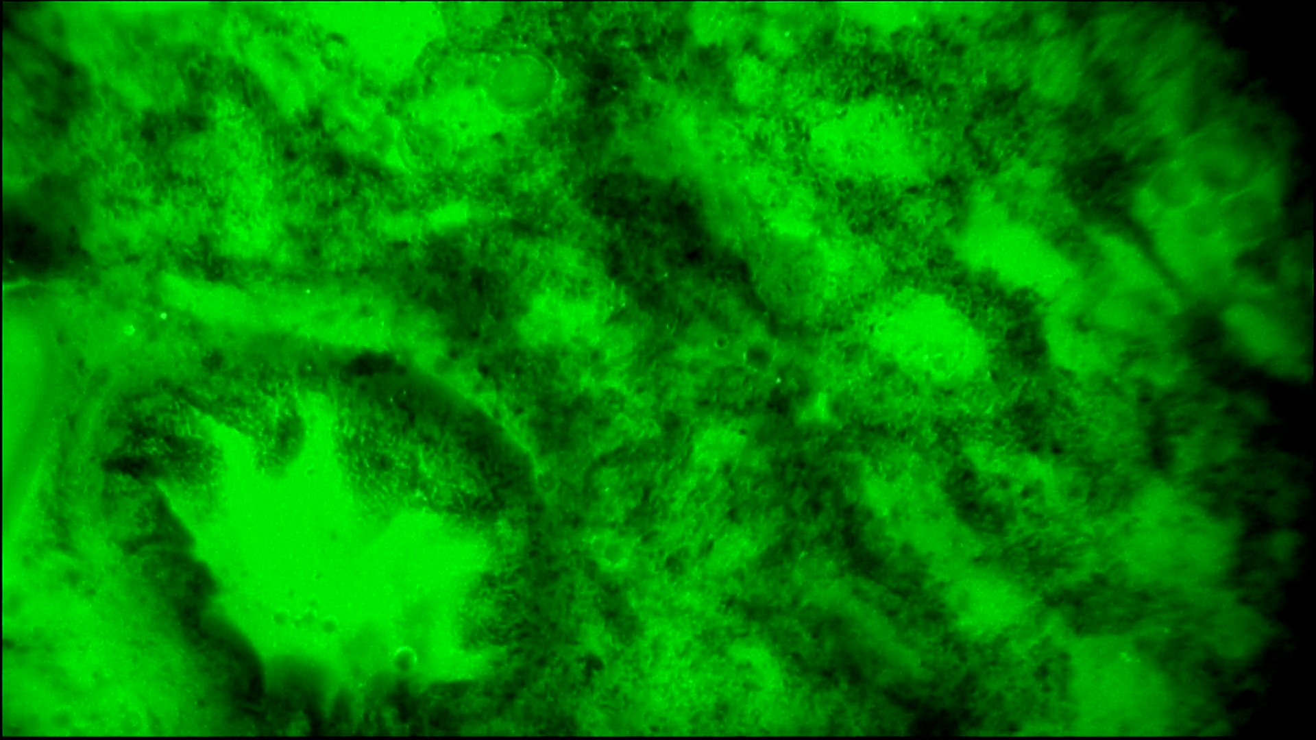
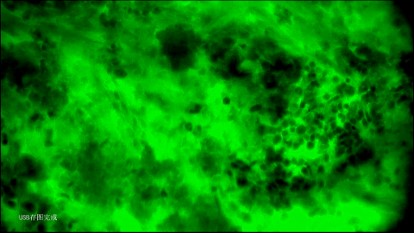
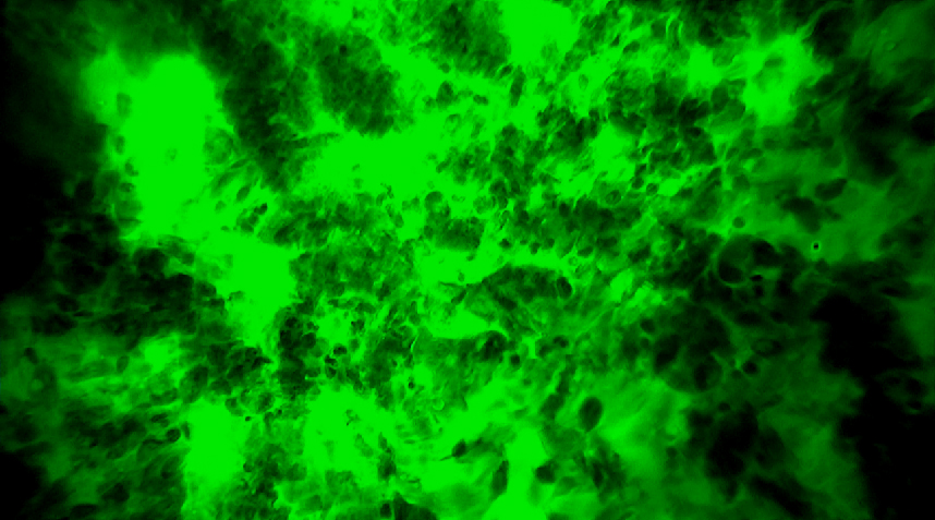
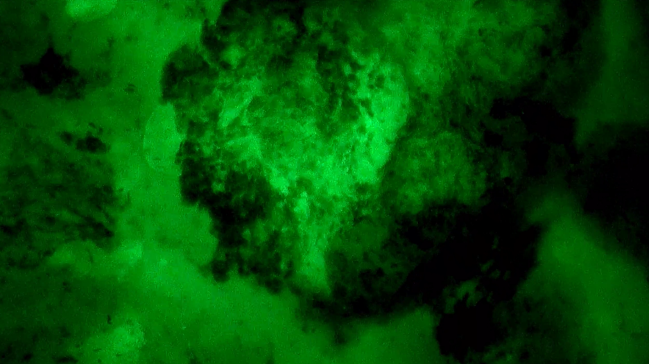
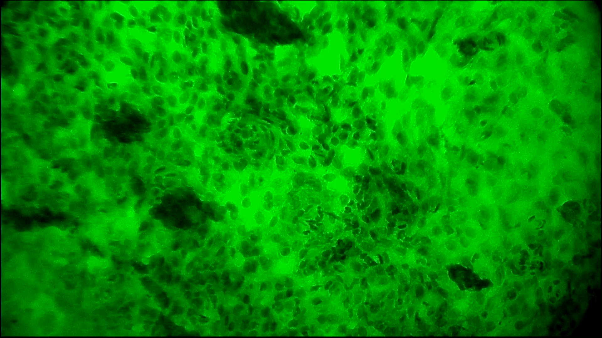
Now, the EndoSCell™ handheld surgical microscope, which is 1,000 times smaller than traditional microscopes, provides real-time, high-definition, dynamic, and artifact-free cellular imaging during surgery. This innovation is expected to expand Mohs surgery to over 80 types of cancers, including gliomas, breast cancer, liver cancer, prostate cancer, ovarian cancer, salivary gland tumors, bladder cancer, and oral cancer.


